Currents, channels, and migraines: The electrical journey of our neurons
- Science and society
on the April 7, 2026
For more than two centuries, scientists have been investigating an astonishing phenomenon: our bodies generate electricity, and our neurons use it to communicate at high speeds.
From Galvani’s frog to modern techniques capable of opening or closing an ion channel with a single flash of light… the history of bioelectricity is full of dramatic twists and turns. At the Valrose Institute of Biology, Guillaume Sandoz’s team is currently exploring a family of channels that is crucial to understanding pain and migraines.
LEAPING FROGS: THE BIRTH OF BIOELECTRICITY
It all began in 1756, in Padua. The physician Leopoldo Caldani applied electrical discharges to muscles and to an isolated heart. And then, a miracle:
it moved! The frog quickly became the star animal of the experiments: hypersensitive, it revealed that nerves react to electricity like tiny living cables.
A few years later, in 1780, Luigi Galvani noticed that a frog’s leg twitched when connected to two metals. He saw this as proof of “animal electricity” produced by the body. But not everyone agreed. Alessandro Volta, for his part, believed that the current came from the metals, not the body. To support his idea, he invented the first electric battery in 1799.
A fruitful disagreement: the debates between Galvani and Volta gave birth to modern electrophysiology.
THE ELECTRICAL LANGUAGE OF NEURONS
Why electricity? Because it’s ultra-fast. Neurons convert information into electrical impulses that travel along nerve fibers in a matter of milliseconds. At the heart of this mechanism: ion channels, tiny gates embedded in the cell membrane. By opening or closing, they allow ions to pass through and generate electrical currents.
Starting in the 1940s, and then in 1960, Alan Hodgkin and Andrew Huxley studied the giant squid axon—so large that an electrode could be inserted into it. Their work, which earned them a Nobel Prize in 1963, described the electrical functioning of the neuron with unprecedented precision.
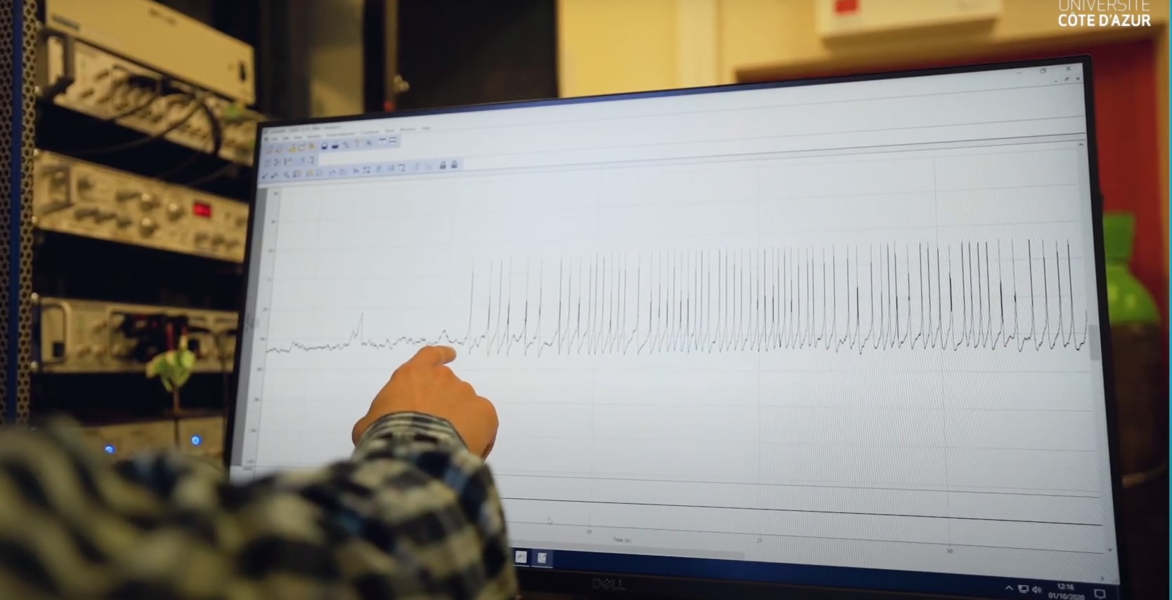
In the late 1970s, the patch-clamp method emerged, was refined, and made it possible for the first time to record the current of a single ion channel. Then in 2003, Rod MacKinnon won the Nobel Prize for the first 3D structure of an ion channel. In 2021, David Julius and Ardem Patapoutian were also honored for the discovery of channels that enable the sensing of heat, cold, or pressure. In a century, our neurons have ceased to be black boxes.
At the Valrose Institute of Biology, Guillaume Sandoz’s team is focusing on a family of channels discovered in 1996: K2P channels, short for “two pore domains.” Their role? To allow potassium ions to escape.
to maintain the electrical calm of neurons. When these gates close, neurons become more sensitive. This increased sensitivity can trigger various conditions, including chronic pain and migraines—the latter affecting women three times more often than men.
In 2010, a study showed that a mutation in the TRESK channel is associated with a familial form of migraine. Nine years later, the Nice-based team demonstrated that certain mutations block not only TRESK but also two of its relatives: TREK1 and TREK2. When these three channels are disabled, neurons become hyper-excitable, and a migraine sets in. But the good news is that by targeting TREK1 and TREK2, normal function can be restored, paving the way for new therapeutic approaches.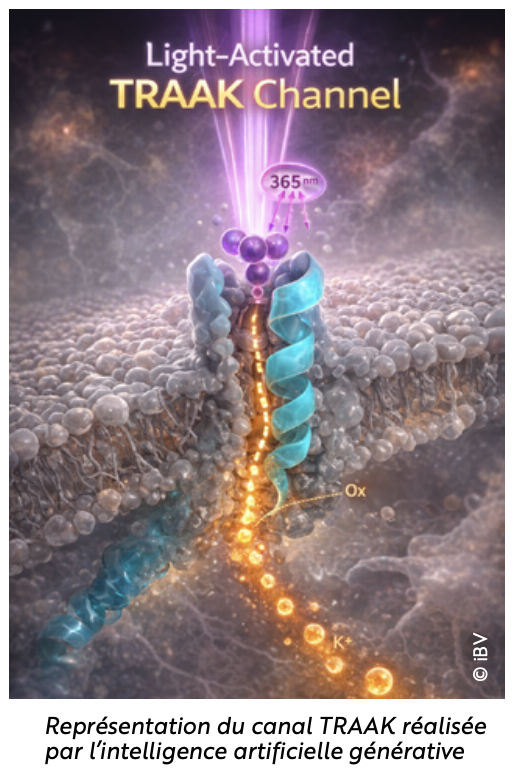
REMOTE-CONTROLLING PAIN: LIGHT AS A NEW MEDICINE
One question remains: how can these channels be precisely controlled? In 2023, researchers modified a molecule and created LAKI. The result: a light-sensitive molecule that changes shape depending on the light it receives. Under violet light, it blocks certain channels, and under green light, the molecule becomes inactive again. In mice, in the dark, nothing happens. But a 20-second flash of violet light is enough to reduce mechanical sensitivity... then the effect disappears. Pain can even be modulated down to the millimeter, just as one would adjust the brightness of a lamp.
And that’s not all: even the C. elegans worm, which has only 302 neurons, changes its behavior under violet light in the presence of LAKI.
More recently, the team demonstrated that, in mice, some of these channels can be directly activated by UV light. In rodents, simple exposure to this light is sufficient to reduce pain. Molecular analyses reveal
that a specific amino acid makes the TRAAK channel sensitive to UV light. If it is replaced, the sensitivity disappears. The light therefore actually activates the channel, hyperpolarizes the neurons... and reduces pain.
A PATHWAY FOR FUTURE PAIN RELIEVERS?
In mice, the pain tolerance threshold is tested by measuring the time it takes for the paw to be withdrawn. Light activation of the channel doubles this time; the mice are therefore less sensitive to pain. This effect lasts for several hours. The effect even surpasses that of certain conventional painkillers like ibuprofen, including for chronic pain. When tested in rats, the method works just as well.
This approach opens up a new avenue: manipulating K2P channels with light to provide precise, reversible, and non-invasive analgesia. A bold idea and promising results that could transform pain management.
If you’d like to learn more about this topic, you can read this article in The Conversation.