Discovery of an endogenous activator of atypical NMDA receptor function
Research
Published on July 24, 2024–Updated on July 24, 2024
Dates
on the June 24, 2024
recherche labo
In a paper published in the journal Neuron in June 2024, Dr. Marie's team at the Institut de Pharmacologie Moléculaire et Cellualire ( Université Côte d'Azur, CNRS), in collaboration with other national and international researchers, identifies the endogenously secreted protein, called AETA, as an essential modulator of the NMDA receptor. The identification of this new AETA-dependent mechanism promises to be a potential therapeutic target for many brain pathologies associated with NMDA receptor dysfunction.
NMDA receptors are essential for most cognitive processes in the central nervous system, as they control the strength of synapses during changes in brain activity. Impaired NMDA receptor function is observed in many brain diseases, including Alzheimer's disease, Parkinson's disease, Huntington's disease, schizophrenia, depression, stroke, epilepsy, autism spectrum disorders, intellectual disability and anti-NMDA receptor encephalitis.
NMDA receptors function in an alternative, atypical mode
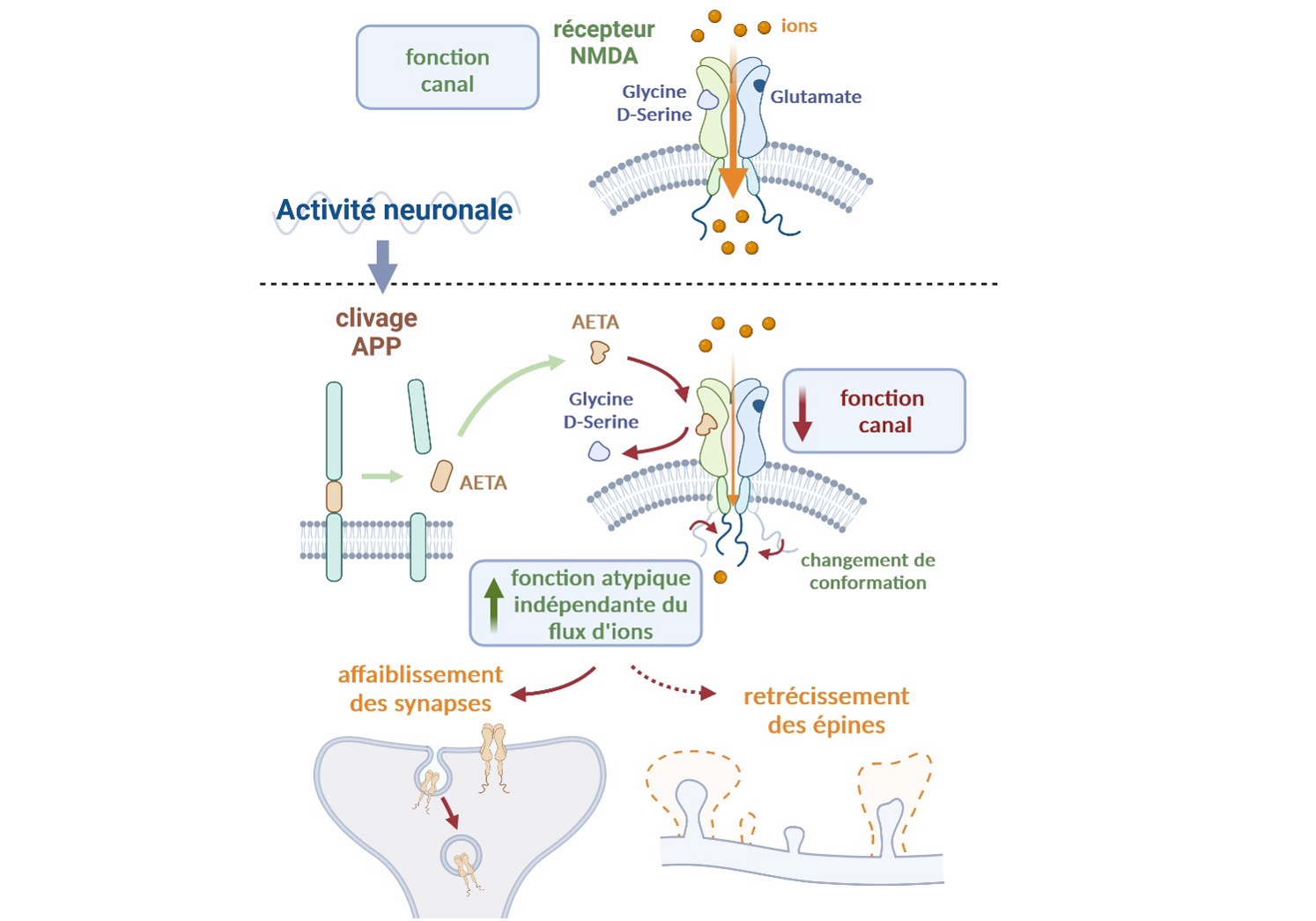
For decades, scientists thought that NMDA receptors functioned essentially as ion channels, requiring the binding of glutamate and the co-agonists glycine or D-serine. This function allows the entry of calcium into synapses to activate a cascade of events necessary for the transmission of information. However, recent evidence suggests that NMDA receptors may also function in an alternative, atypical mode. This mode does not require the passage of ions through the channel, but rather a conformational change leading to an activity independent of ion flow. This atypical activity is linked to the weakening of synapses, a crucial process for regulating connectivity between neurons. This dual mode of activity remains enigmatic. Can the NMDA receptor switch rapidly between its two modes of activity? If so, how? Does this have an impact on cognition? Is it dependent on neuronal activity?
How does AETA act at the synaptic level?
In this article, the authors describe AETA as a new molecular player that controls this switching. The existence of AETA (also known as A) was first described by Willem et al. in 2015, in collaboration with Dr. Marie's team (Willem et al. Nature 2015). AETA is cleaved from the amyloid precursor protein- (APP), a protein strongly implicated in Alzheimer's disease, and then secreted into the brain. The authors had previously shown that direct contact with small quantities of soluble AETA immediately reduced neuronal activity in the central nervous system, but the mechanism of action was unknown.
Now, an international consortium of scientists has worked with Dr Marie to explain how AETA acts at the level of synapses, using electrophysiological recordings and functional imaging. With elegant experiments, they demonstrate that AETA directly and specifically inhibits the ion channel function of the NMDA receptor in competition with co-agonists (glycine/D-serine). AETA binding leads to a conformational change in the receptor, associated with an increase in the receptor's alternative atypical activity, which does not require ionic flux. AETA binding promotes this atypical activity of the NMDA receptor, leading to weakening of synapses and shrinkage of dendritic spines, the structural units hosting synapses. No other known endogenous brain molecule possesses this unique property of acting as a key unlocking this atypical function.
The authors also created new mouse models lacking or over-expressing AETA to demonstrate the necessity ofAETA in the control of NMDA receptor signaling, synapse impairment and NMDA receptor-dependent memory formation. Finally, using chemogenetics, they show that AETA production is increased when neurons are activated in vivo, providing further evidence that regulation of AETA-dependent NMDA receptor activity is an essential physiological mechanism for cerebral information processing. The next steps will be to determine whether this novel endogenous mechanism is compromised in the various brain diseases involving the NMDA receptor. This exploration will offer great opportunities for seeking innovative strategies to treat the many brain disorders linked to this receptor.
schema H mari IPMC
Figure: the small protein AETA, which is cleaved from amyloid-β precursor protein (APP) during increased neuronal activity, competes with D-serine/glycine to inhibit NMDA receptor channel function. AETA binding leads to a conformational change in the receptor. AETA-dependent modulation of the receptor induces synapse weakening and dendritic spine narrowing, two functions of the NMDA receptor linked to its atypical mode of activity independent of ion flow. Diagram created by Biorender.com.
When browsing Université Côte d'Azur website and Université Côte d'Azur components websites by profile ("I am" menu), informations may be saved in a "Cookie" file installed by Université Côte d'Azur on your computer, tablet or mobile phone.This Cookie file contains informations, such as a unique identifier, the name of the portal, and the chosen profile. This Cookie file is read by its transmitter. During its 12-month validity period, it allows to recognize your terminal and to propose the chosen profile as your default home page.
You have accepted the deposit of profile information cookies in your navigator.
You have declined the deposit of profile information cookies in your navigator.
"Do Not Track" is enabled in your browser. No profiles information will be collected.