KEY POINTS
-
Organoids are miniature organ-like structures that replicate certain characteristics of organs.
-
A research team is working on brain organoids to study the behavior of glioblastoma cells (brain cancer).
-
Improving these brain models raises ethical questions: could they become “mini-brains”?
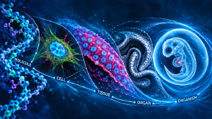
For a long time, the processes governing human embryonic development during early fetal stages were difficult to study, for both technical and ethical reasons. Researchers therefore had to turn to experimental models such as animals, which are certainly useful but sometimes far removed from human physiology. However, a revolution has profoundly transformed developmental biology in recent decades: the advent of organoids. These are organ prototypes—from the Greek organon (“organ”) and -oid (“resembling”) —that is, miniaturized three-dimensional structures derived from pluripotent stem cells cultured in the laboratory, capable of reproducing certain structural and functional characteristics of human organs (notably the intestine, brain, retina, liver, kidney, pancreas, lung, and stomach).
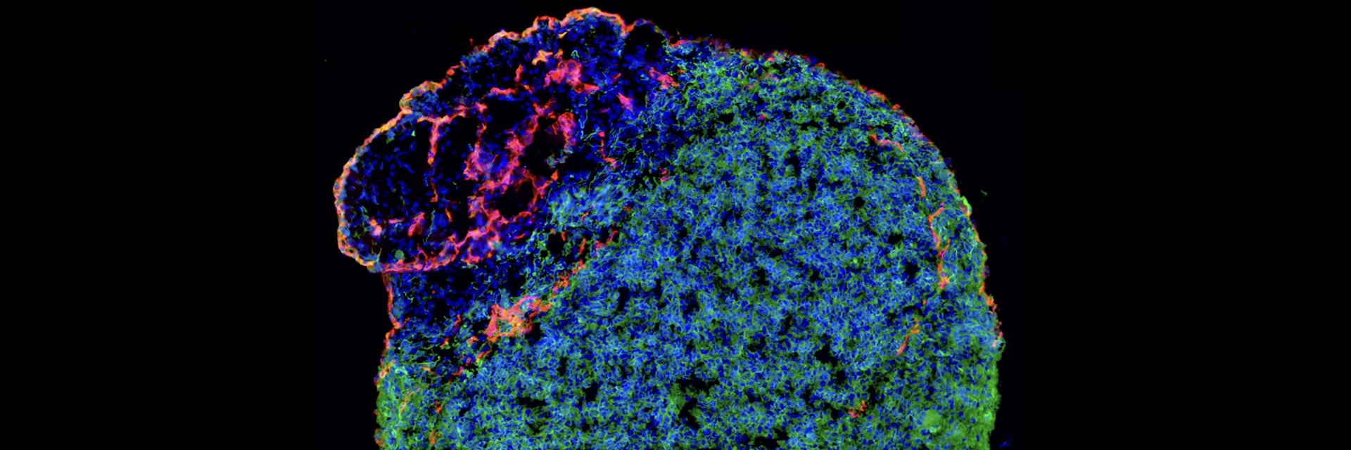
In the case of the brain, brain organoids are 3D aggregates of neuronal tissue, within which progenitor cells generate neurons. These neurons differentiate, arrange themselves into rudimentary layers, and establish connections, forming simplified networks. These networks exhibit spontaneous electrical activity that, within certain limits, resembles that observed during early human brain development (first trimester). This is therefore a remarkable strategy for exploring otherwise inaccessible windows of human development.
Observing Brain Development in a Petri Dish
When I joined Dr. Michèle Studer’s laboratory at the Valrose Institute of Biology (IBV) at the Université Côte d’Azur, I began to develop and use this approach to study brain development. It is an extraordinary tool that allows us to model stages of brain development in vitro, in a Petri dish. In the case of genetic manipulations, it helps us understand how these particularly delicate early processes are disrupted in conditions leading to neurodevelopmental disorders, such as autism spectrum disorders, intellectual disability, or certain forms of epilepsy. Simpler and more accessible than a human brain, these systems facilitate experimental work. For example, to study the role of a gene involved in brain development, one need only compare organoids derived from unmodified stem cells—used as controls—with organoids derived from genetically modified stem cells, for example, through mutation or gene knockdown.
However, a limitation quickly becomes apparent: these organoids remain highly simplified compared to a human brain. While simplification is useful, it must not obscure essential properties of the organ under study. The scientific community has recognized a fundamental aspect: neurodevelopmental disorders almost never affect just a single cell type or a single region of the brain. They often involve multiple cell types, multiple regions, and the interactions between these cells. It is in this context that a new approach has emerged: increasing the complexity of organoids to better reflect the organization of the brain in vivo.
Increasing the Complexity of Organoids
One strategy involves separately generating organoids with distinct regional identities—for example, corresponding to the dorsal and ventral parts of the brain—and then fusing them together. These systems, called “assembloids,” notably replicate the natural migration of inhibitory neurons from the ventral region to the dorsal region, where they integrate into neural circuits. This type of model thus allows for the simplified study of key developmental phenomena, such as neuronal migration and the balance between excitation and inhibition, which are often disrupted in neurodevelopmental disorders such as certain forms of genetically caused epilepsy. Even more complex systems have been developed, combining several regions of the nervous system and even peripheral tissues. For example, multistep circuits connecting brain-like structures, the spinal cord, and muscles have been reconstructed, making it possible to observe muscle contractions in response to neural stimulation.
Other approaches involve the use of morphogens—molecules that mimic the chemical signals guiding brain organization during embryonic development. These signals provide cells with “spatial coordinates,” telling them where to position themselves and how to organize, thereby allowing for greater control over the organization and “regionalization” of organoids—that is, the formation of multiple regions within a single organoid—without the need to develop them separately and then fuse them together. These models remain highly simplified but are gradually approaching the complexity of real biological systems.
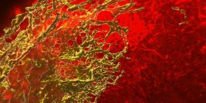
Beyond neurons, the brain comprises a wide variety of cell types. Among them, endothelial cells form the blood vessels that supply the brain tissue and contribute to the blood-brain barrier, an essential interface that regulates exchanges between the blood and the brain and protects the brain from external threats. Interactions between neurons and blood vessels play a major role in many diseases. For example, in glioblastoma, tumor cells can migrate along blood vessels and compromise the integrity of the blood-brain barrier. We have recently developed brain organoids enriched with endothelial cells, enabling us to reproduce capillary-like structures within neuronal tissue. Although there is no blood circulation in these models, the presence of vascular structures significantly increases cellular complexity and heterogeneity, paving the way for the study of new biological interactions.
Gaining a Better Understanding of Glioblastoma
In collaboration with the laboratory of Dr. Thierry Virolle, a research director at the same institute, these organoids were used to study the behavior of glioblastoma cells, particularly their preferential migration toward neuronal or vascular structures depending on their characteristics. By increasing the system’s complexity, we were able to better understand the dynamics of tumor invasion, particularly by identifying molecular switches capable of redirecting glioblastoma cells toward different cell types. Ultimately, complex organoids represent a new generation of biological models, more closely resembling the actual organization of the organs they replicate.
But how far can we push this complexity? This question is now at the heart of discussions in the field, particularly from an ethical standpoint. Organoids already offer an ethically sound alternative to animal experimentation, while being more representative of human physiology. However, as they become more complex and reach more advanced stages of maturation—comparable to certain later stages of fetal development, particularly during the second trimester—new questions arise. Are they still merely experimental models, or biological systems endowed with more advanced properties? Will complex brain organoids one day be capable of developing forms of activity comparable to those of a miniaturized human brain? While this prospect remains distant for current organoids, it is nevertheless beginning to be seriously discussed within the scientific community.
Michele Bertacchi, Inserm Researcher, Valrose Institute of Biology, Université Côte d'Azur, and Michèle Studer, Inserm Research Director of Exceptional Class (DRCE), Université Côte d’Azur